Welcome to the Amira-Avizo Software Use Case Gallery
Below you will find a collection of use cases of our 3D data visualization and analysis software. These use cases include scientific publications, articles, papers, posters, presentations or even videos that show how Amira-Avizo Software is used to address various scientific and industrial research topics.
Use the Domain selector to filter by main application area, and use the Search box to enter keywords related to specific topics you are interested in.
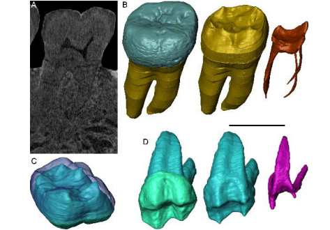
Exploring hominin and non-hominin primate dental fossil remains with neutron microtomography
Fossil dental remains are an archive of unique information for paleobiological studies. Computed microtomography based on Xray microfocus sources (X-µCT) and Synchrotron Radiation (SR-µCT) allow subtle quantification at the micron and sub-micron scale of the meso- and microstructural signature imprinted in the mineralized tissues, such as enamel and dentine, through highresolution “virtual histology”. Nonetheless, depending on the degree of alterations undergone during fossiliza... Read more
Clément Zanolli, Laboratory AMIS, UMR 5288, University of Toulouse III - Paul Sabatier, France, and al.
Collagen-Based Matrices for Osteoconduction: A Preclinical In Vivo Study
The aim of this study was to evaluate the influence of additional hydroxyapatite (HA) in collagen-based matrices (CM) and membrane placement on bone formation in calvarial defects.
Critical size defects in the calvaria of 16 New Zealand White Rabbits were randomly treated with CM or mineralized collagen-based matrices (mCM). Half of the sites were covered with a collagen membrane. Animals were euthanized after 12 weeks of healing. The samples were studied by micro-CT and histology. New... Read more
Hiroki Katagiri, Yacine El Tawil, Niklaus P. Lang, Jean-Claude Imber, Anton Sculean, Masako Fujioka-Kobayashi and Nikola Saulacic
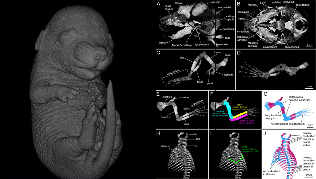
For decades, clearing and staining with Alcian Blue and Alizarin Red has been the gold standard to image vertebrate skeletal development. Here, we present an alternate approach to visualise bone and cartilage based on X-ray microCT imaging, which allows the collection of genuine 3D data of the entire developing skeleton at micron resolution.
Our novel protocol is based on ethanol fixation and staining with Ruthenium Red, and efficiently contrasts cartilage matrix, as demonstrated in wh... Read more
Simone Gabner, Peter Böck, Dieter Fink, Martin Glösmann, Stephan Handschuh
Progressive transformation of the otic placode into the functional inner ear during gestational development in humans leads to the acquisition of hearing perception via the cochlea and balance and spatial orientation via the vestibular organ.
Using a correlative approach involving micro-computerized tomography (micro-CT), transmission electron microscopy and histological techniques we were able to examine both the morphological and cellular changes associated with human inner ear devel... Read more
Lejo Johnson Chacko, David Wertjanz, Consolato Sergi, Jozsef Dudas, Natalie Fischer, Theresa Eberharter, Romed Hoermann, Rudolf Glueckert, Helga Fritsch, Helge Rask-Andersen, Anneliese Schrott-Fischer & Stephan Handschuh
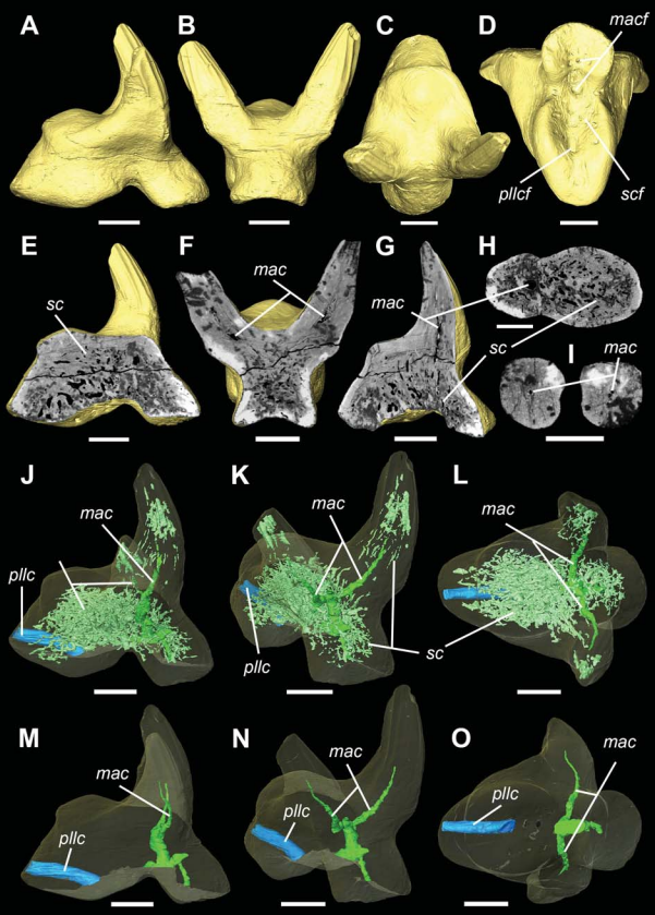
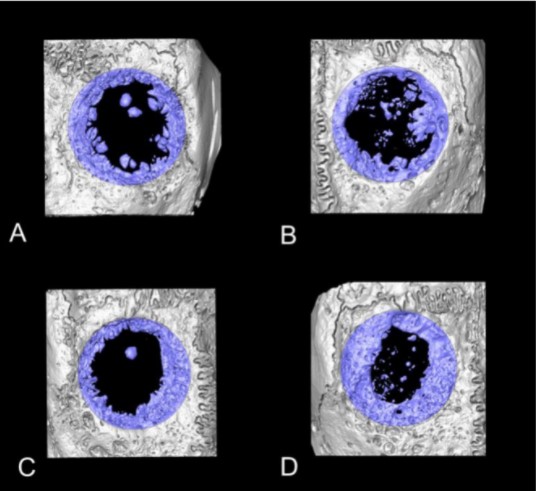
Vascular structure of the earliest shark teeth
Here we use synchrotron tomography to characterise dental vasculature in the oldest known tooth-bearing sharks, Leonodus carlsi Mader, 1986 and Celtiberina maderi Wang, 1993. Three dimensional reconstruction of the vascular system and microstructure of both taxa revealed a complex and dense network of canals, including horizontal, ascending and secondary bifurcated canals, as well as histological features consistent with an osteodont histotype. However, L. carlsi and C. maderi also exhibit si... Read more
Carlos Martinez-Perez, Alba Martin-Lazaro, Humberto G Ferron, Martina Kirstein, Philip C.J. Donoghue, Hector Botella
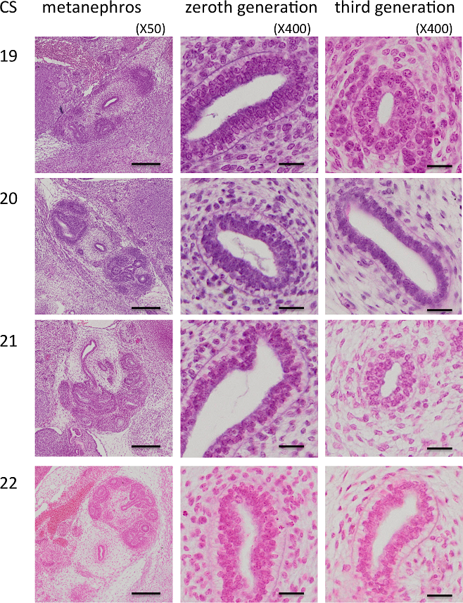
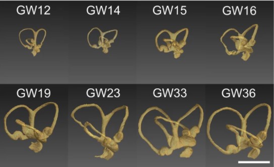
Branching morphogenesis of the urinary collecting system in the human embryonic metanephros
An elaborate system of ducts collects urine from all nephrons, and this structure is known as the urinary collecting system (UCS). This study focused on how the UCS is formed during human embryogenesis. Fifty human embryos between the Carnegie stage (CS) 14 and CS23 were selected from the Kyoto Collection at the Congenital Anomaly Research Center of Kyoto University, Japan. Metanephroses, including the UCS, were segmented on serial digital virtual histological sections. Three-dimensional imag... Read more
Hana Ishiyama, Aoi Ishikawa, Haruka Kitazawa, Sena Fujii, Jun Matsubayashi, Shigehito Yamada, Tetsuya Takakuwa
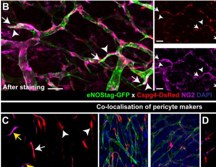
Endothelial cells and pericytes are integral cellular components of the vasculature with distinct interactive functionalities. To study dynamic interactions between these two cells we created two transgenic animal lines. A truncated eNOS (endothelial nitric oxide synthase) construct was used as a GFP tag for endothelial cell evaluation and an inducible Cre-lox recombination, under control of the Pdgfrb (platelet derived growth factor receptor beta) promoter, was created for pericyte assessmen... Read more
Ann L. B. Seynhaeve, Douwe Oostinga, Rien van Haperen, Hanna M. Eilken, Susanne Adams, Ralf H. Adams & Timo L. M. ten Hagen
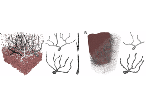
Three-dimensional virtual histology of human cerebellum by X-ray phase-contrast tomography
To quantitatively evaluate brain tissue and its corresponding function, knowledge of the 3D cellular distribution is essential. The gold standard to obtain this information is histology, a destructive and labor-intensive technique where the specimen is sliced and examined under a light microscope, providing 3D information at nonisotropic resolution. To overcome the limitations of conventional histology, we use phase-contrast X-ray tomography with optimized optics, reconstruction, and image an... Read more
Mareike Töpperwien, Franziska van der Meer, Christine Stadelmann, and Tim Salditt